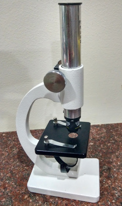
Microscope
Last week our class started with discussion about what make up our
bodies, plants and all living beings. Kids knew the answer as cells.
So, the question was
1. Can we see these cells with naked eyes?.
2. So, if we have some illness , how does a doctor understand what kind of treatment to be done?.
3. If the doctor advises for blood test, what do the labs do with the blood sample?.
Depending on the type of the blood test, to see these cells, bacteria etc we need special instrument called ' microscope'.
Microscope
Microscopy is the science of investigating small objects and structures using special instrument called 'Microscope'. The three main different types of microscope are dissection, compound and electron microscope. By using microscopes
scientists were able to discover the existence of microorganisms, study
the structure of cells, and see the smallest parts of plants, animals,
and fungi.
The microscope that we are using is compound microscope. Compound microscopes are light illuminated and the images seen are two dimensional. They were eager to know about the different parts of the microscope, their purpose and slides with specimen. They checked out slides with onion skin, mosquito etc. They each got to see it by themselves.We then made our temporary slides of leaf, hair, sugar, salt crystals.
One of the students had brought a dead fly because he had checked with me earlier to find out what activity I was planning to teach. It sure was good to see kids coming prepared.
Curious to see what is on their skin, they even tried looking at their fingers through the microscope which was too thick for it to be visible. They were confused and wanted to know the reason.
Do you know why you can not see your fingers directly under the microscope?
You can see the objects under the microscope only when when light passes through them. Opaque objects are not visible. So, we then made a temporary slide of their finger print.
To see the leaf structure we also made a slide of a leaf by cutting the cross section of the leaf. It was easy for the students to relate to because they had studied about the stomata, epidermis etc. To see it through the microscope was impressive for them.
Please note that to see clearly through the microscope, you need to keep the whole set up outside in natural light . It was exciting for kids to observe through the microscope.


Comments
Post a Comment
Share your thoughts here: